The year's best microscopic images reveal an eerie and breathtaking world that most of us never see

Advertisement
The year's best microscopic images reveal an eerie and breathtaking world that most of us never see
  • The Nikon Small World contest highlights the best microscope images taken each year.
  • This year, the contest received more than 2,000 entries from people in 90 countries.
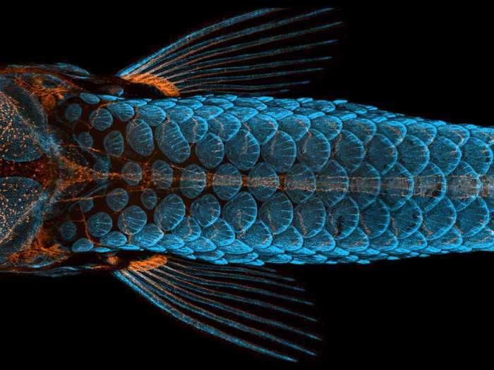
  • The winning image of 2020, of a young zebrafish threaded with neon-orange lymphatic vessels, reveals the species could help researchers find treatments for brain diseases.
  • Other winning images include a rainbow snail tongue, a curled-up chameleon embryo, and the intricate mesh of a nylon stocking.

If there's one thing we've learned from the coronavirus pandemic, it's a newfound respect for the very, very small.

But as dangerous as the microscopic universe can seem, it's also rich with stunning shapes, brilliant colors, and often mysterious biological and physical processes.

In that spirit, Nikon has released the winners of Small World: the camera company's 46th microscopy, or microscope photography, competition.

Judges Dylan Burnette, Christophe Leterrier, Samantha Clark, Sean Greene, and Ariel Waldman reviewed more than 2,000 entries from scientists, artists, and hobbyists from 90 countries around the world. All have to be taken with microscopes using optical imaging techniques. They include a rainbow snail tongue, a self-replicating worm, and – appropriate for the Halloween season – a spooky-looking bat embryo skeleton.

Here are the best microscope pictures of the year and what they show.
Advertisement

First place went to an image that revealed a stunning truth about zebrafish.

First place went to an image that revealed a stunning truth about zebrafish.
Dorsal view of bones and scales (blue) and lymphatic vessels (orange) in a juvenile zebrafish. Daniel Castranova, Dr. Brant Weinstein & Bakary Samasa/Nikon Small World

The photo isn't just gorgeous and technically perfect: It also led researchers to a groundbreaking discovery.

The orange in the photo represents the zebrafish's lymphatic system, the network of vessels that carry white blood cells through the body. Previously, scientists thought that only mammals had lymphatic vessels inside their skulls. But with this photo, photographers Daniel Castranova and Bakary Samsara at the National Institutes of Health show that zebrafish have those vessels too.

Because fish are so much easier to raise and conduct experiments on than mammals, this discovery could revolutionize research around treatments for brain-related diseases, like Alzheimer's disease and brain cancer.

An exquisite image of a clownfish embryo over time, from hours after fertilization to hours before hatching, took second place.

An exquisite image of a clownfish embryo over time, from hours after fertilization to hours before hatching, took second place.
Embryonic development of a clownfish (Amphiprion percula) on days 1, 3 (morning and evening), 5, and 9. Daniel Knop/Nikon Small World
Advertisement

A rainbow image showing the ridged tongue of a freshwater snail came in third.

A rainbow image showing the ridged tongue of a freshwater snail came in third.
Tongue (radula) of a freshwater snail. Dr. Igor Siwanowicz/Nikon Small World

Here are the rest of the 20 winners:

4. Stringy filaments and spores of a soil fungus.

4. Stringy filaments and spores of a soil fungus.
Multi-nucleate spores and hyphae of a soil fungus (arbuscular mycorrhizal fungus). Dr. Vasileios Kokkoris, Dr. Franck Stefani & Dr. Nicolas Corradi/Nikon Small World
Advertisement

5. The feathery, huge-eyed profile of a bogong moth.

5. The feathery, huge-eyed profile of a bogong moth.
A bogong moth. Ahmad Fauzan/Nikon Small World

6. The anther – or pollen-containing section – of a Hebe shrub's flower.

6. The anther – or pollen-containing section – of a Hebe shrub's flower.
Hebe plant anther with pollen. Dr. Robert Markus & Zsuzsa Markus/Nikon Small World
Advertisement

7. A thicket of tiny microtubules within a cell, surrounding the egg-shaped nucleus.

7. A thicket of tiny microtubules within a cell, surrounding the egg-shaped nucleus.
Microtubules (orange) inside a cell. Nucleus is shown in cyan. Jason Kirk/Nikon Small World

8. A spooky chameleon embryo.

8. A spooky chameleon embryo.
Chameleon embryo (autofluorescence). Dr. Allan Carrillo-Baltodano & David Salamanca/Nikon Small World
Advertisement

9. Connections between brain cells that look a bit like lightning.

9. Connections between brain cells that look a bit like lightning.
Connections between hippocampal neurons (brain cells). Jason Kirk & Quynh Nguyen/Nikon Small World

10. Daphnia magna, a crustacean that's smaller than a pencil eraser.

10. Daphnia magna, a crustacean that's smaller than a pencil eraser.
Daphnia magna (Phyllopoda). Ahmad Fauzan/Nikon Small World
Advertisement

11. Those aren't mutant skeleton fingers – they're microscopic depictions of red algae.

11. Those aren't mutant skeleton fingers – they're microscopic depictions of red algae.
Red algae. Dr. Tagide deCarvalho/Nikon Small World

12. Human hair tied in a knot.

12. Human hair tied in a knot.
Human hair. Robert Vierthaler/Nikon Small World
Advertisement

13. A dreamlike image of crystals formed after heating a solution of amino acids used in muscle building.

13. A dreamlike image of crystals formed after heating a solution of amino acids used in muscle building.
Crystals formed after heating an ethanol and water solution containing L-glutamine and beta-alanine. Justin Zoll/Nikon Small World

14. The side view of an intrepid, iridescent leaf roller weevil.

14. The side view of an intrepid, iridescent leaf roller weevil.
Leaf roller weevil (Byctiscus betulae) lateral view. Özgür Kerem Bulur/Nikon Small World
Advertisement

15. A chain of developing Chaetogaster diaphanus worms, which reproduce asexually by multiplying themselves.

15. A chain of developing Chaetogaster diaphanus worms, which reproduce asexually by multiplying themselves.
Chain of daughter individuals from the asexually reproducing annelid species Chaetogaster diaphanus. Dr. Eduardo Zattara & Dr. Alexa Bely/Nikon Small World

16. The intricate net of a nylon stocking, magnified nine times.

16. The intricate net of a nylon stocking, magnified nine times.
Nylon stockings. Alexander Klepnev/Nikon Small World
Advertisement

17. A pastel portrait of a water boatman, a type of aquatic insect with oar-like legs.

17. A pastel portrait of a water boatman, a type of aquatic insect with oar-like legs.
Ventral view of an immature water boatman. Anne Algar/Nikon Small World

18. The fuzzy outline of an Atlas moth wing.

18. The fuzzy outline of an Atlas moth wing.
Atlas moth wing. Chris Perani/Nikon Small World
Advertisement

19. The perfectly round cell wall of Arachnoidiscus sp., a type of microscopic floating algae.

19. The perfectly round cell wall of Arachnoidiscus sp., a type of microscopic floating algae.
Silica cell wall of the marine diatom Arachnoidiscus sp. Dr. Jan Michels/Nikon Small World

20. A Halloween-esque image of a fruit bat embryo skeleton.

20. A Halloween-esque image of a fruit bat embryo skeleton.
Skeleton preparation of a short-tailed fruit bat embryo (Carollia perspicillata). Dr. Dorit Hockman & Dr. Vanessa Chong-Morrison/Nikon Small World
Advertisement