The year's best microscope photos reveal a hidden world, from a pregnant flea to crystals in a dinosaur bone

Advertisement
The year's best microscope photos reveal a hidden world, from a pregnant flea to crystals in a dinosaur bone
  • Nikon's Small World competition recognizes the art and science of microscope photography.
  • This year's winning images reveal firing neurons, the vessels of leaves and eyeballs, and colorful crystals inside a dinosaur bone.

Everyday things look quite different under a microscope - including the leaf of a southern live oak tree, pictured above.

Jason Kirk, director of a microscope-imaging facility at Baylor College of Medicine, likes to tinker with microscopes and use them to photograph things from his backyard. To create the above image, he illuminated a leaf's small-scale structures with a customized microscope. Kirk captured about 200 images then stacked them on top of each other to create this vibrant portrait.

The result shows the leaf's trichomes - little outgrowths that protect the plant - in white. Pores that help the plant regulate gas flows, called stomata, appear in purple. Vessels that carry water through the leaf pop out in blue-green.

That technical handiwork led Kirk to win first place in this year's Nikon Small World competition, a contest the camera company launched in 1974 to recognize achievements in microscope photography, also known as microscopy.

"Nikon Small World was created to show the world how art and science come together under the microscope. This year's first-place winner could not be a better example of that blend," Eric Flem, a communications manager at Nikon Instruments, said in a press release.

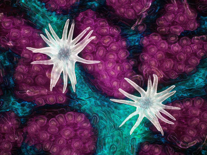
For the 2021 competition, Nikon received nearly 1,900 entries from 88 countries. A panel of judges selected 20 winners and recognized 80 other images for distinction or honorable mention. Some of the photos, like Kirk's, show familiar things in a totally new light. Others reveal organisms all around us that we never see.

Here are 16 other striking photos from the competition.

Advertisement

An image of 300,000 networking neurons inside a microchip took second place in the contest. A bridge of axons - the long fibers that allow neurons to communicate - splits the two groups of neurons.

An image of 300,000 networking neurons inside a microchip took second place in the contest. A bridge of axons - the long fibers that allow neurons to communicate - splits the two groups of neurons.
Esmeralda Paric & Holly Stefen/Nikon Small World

This profile of a hog louse snagged third place. It reveals the tiny bug's leg, claw, and trachea.

This profile of a hog louse snagged third place. It reveals the tiny bug's leg, claw, and trachea.
Frank Reiser/Nikon Small World
Advertisement

Other notable images also show minuscule critters up close, like this red forest ant.

Other notable images also show minuscule critters up close, like this red forest ant.
Dr. Fred Terveer/Nikon Small World

This headshot of a tick reveals some hidden color.

This headshot of a tick reveals some hidden color.
Dr. Tong Zhang & Dr. Paul Stoodley/Nikon Small World
Advertisement

Some of the images zoom in much closer on facets of small creatures - this image shows a single neuron from a rat embryo.

Some of the images zoom in much closer on facets of small creatures - this image shows a single neuron from a rat embryo.
Paula Diaz/Nikon Small World

Rodents and their inner workings are common microscopy subjects. This image shows blood vessels inside the retina of a mouse's eye.

Rodents and their inner workings are common microscopy subjects. This image shows blood vessels inside the retina of a mouse's eye.
Jason Kirk & Carlos P. Flores Suarez/Nikon Small World
Advertisement

Water fleas are also regular microscopy models. This one is pregnant with embryos.

Water fleas are also regular microscopy models. This one is pregnant with embryos.
Jan van IJken/Nikon Small World

Another photo from this year's contest shows brine shrimps eggs after the babies had hatched.

Another photo from this year's contest shows brine shrimps eggs after the babies had hatched.
Waldo Nell/Nikon Small World
Advertisement

Some images get so close to familiar things that they become unrecognizable. This is a vein and scales on a butterfly wing.

Some images get so close to familiar things that they become unrecognizable. This is a vein and scales on a butterfly wing.
Sébastien Malo/Nikon Small World

Putting cotton fabric under a microscope showed how the fibers weave together. The yellow circles are a few stray grains of pollen that got caught.

Putting cotton fabric under a microscope showed how the fibers weave together. The yellow circles are a few stray grains of pollen that got caught.
Dr. Felice Placenti/Nikon Small World
Advertisement

This may look like some kind of ancient tablet, but it's actually a crystal of table salt.

This may look like some kind of ancient tablet, but it's actually a crystal of table salt.
Saulius Gugis/Nikon Small World

Some photographers did find ancient subjects, though, like this 40-million-year-old gnat preserved in amber.

Some photographers did find ancient subjects, though, like this 40-million-year-old gnat preserved in amber.
Levon Biss/Nikon Small World
Advertisement

This dinosaur bone was infiltrated by silica-rich groundwater that turned its insides to agate - a colorful, crystallized quartz.

This dinosaur bone was infiltrated by silica-rich groundwater that turned its insides to agate - a colorful, crystallized quartz.
Norm Barker/Nikon Small World

Slime mold makes a lot of appearances in microscopy, since it forms otherworldly outgrowths in different shapes. This little mold is magnified 10 times.

Slime mold makes a lot of appearances in microscopy, since it forms otherworldly outgrowths in different shapes. This little mold is magnified 10 times.
Alison Pollack/Nikon Small World
Advertisement

One photographer kept it simple and beautiful, capturing a furled portion of a pagoda flower bud.

One photographer kept it simple and beautiful, capturing a furled portion of a pagoda flower bud.
Jose R. Almodovar/Nikon Small World

Another took a look at a sea anemone's mouth and tentacles by lighting up its neurons. The resulting image offers a kind of galactic portrait of the invisible biology around us.

Another took a look at a sea anemone's mouth and tentacles by lighting up its neurons. The resulting image offers a kind of galactic portrait of the invisible biology around us.
Ruohan Zhong/Nikon Small World
Advertisement